AMD (Age Related Macular Degeneration )
Table of Content
Macular degeneration is connected with age and may be described as 21st blindness epidemic and there are 500000 new cases yearly .It is estimated that in Poland the number of new exudative AMD cases amounts to 20000 yearly.
Understanding AMD
AMD is a disease of eye retina. The retina is a thin paper- like tissue cushioning the rear part of eye interior. Photosensitive retina cells are responsible for converting light into electric impulses, which are sent by optic nerve to the brain for interpretation. In the very centre of retina there is the most important area, the macula, commonly called the spot, which consists of highest density photosensitive cells- stamens responsible for acute and detailed vision when, for example, when we recognize people` s faces , read or drive a car, etc.
In patients with AMD, the stamens in the area of macula begin to decay ( atrophy ), which causes the appearance of spots in eyeshot and vision is distorted.
Kinds of AMD
There are two different kinds of AMD : dry and wet / exudative /
- Dry AMD is the commonest form of macular degeneration . In the course of disease , retina metabolism products, known as druses, accumulate under the layer of retina photoreceptors between retina pigment epithelium /RPE/ and Bruch membrane, which support retina. The druses often appear in the eyes of elderly people, however, the increase of size and number of those deposits is the first symptom of AMD. In time , the druses cause damages and decay of photoreceptors, which lead to seizure and disturbances in the ability of macula central vision. This process does not cause any pain, and in the first stages of disease may notice only slight worsening of vision. However , when more and more photosensitive cells decay, central vision worsens drastically. In more advanced cases, dry AMD may cause substantial vision loss, seriously affecting quality of life.
AMD is the main reason of vision loss In people over 60 years of age.
- Wet / exudative/ AMD
Wet AMD appears when faulty blood vessels start to develop under retina, and that leads to damage and decay of both photoreceptors and retina pigment epithelium /RPE /, which feed the photoreceptors. At first, appear disruptions Bruch membrane, usually around accumulated druses, and new vessels begin to develop and grow into the retina That process is called neovascularisation. Those vessels are responsible for distortion on retina surface ; they are very delicate and break easily, which may result in haemorrhage leading to irreversible retina damage like fibrous scars. All these distortions cause impaired central vision, which may be not sharp. Image may be blurred, crooked and obscured by spots of different sizes and brightness.
FACTS:
Both dry and wet AMD may appear at the same time, even in the same eye.
Dry AMD may be in one eye and wet AMD in the other.
Development of disease may be slow or fast.
Wet AMD develops always much faster than the dry, and in short time , even in a few weeks may lead to irreversible loss of vision.
Dry AMD may lead to loss of vision, not becoming wet.
It is also possible that dry AMD may become wet, already in early stage.
FACTS:
About 8-10% , and according to other sources 20% of AMD patients get the wet AMD. However, in 90% of AMD patients blindness results from improper formation of blood vessels in neovascularisation / CNV /. In most cases. In most cases the progress of disease is so fast that within two years the affected eye practically loses vision.
Worsening and loss of vision may happen very fast, even in days or weeks .Wet AMD constitutes about 10 % to 20% of all AMD cases , but is the reason of 80% to 90 % of practical blindness caused by AMD. All cases of wet AMD are regarded as advanced cases.
What are the symptoms of AMD?
One must not disregard any vision disorders, or wait till they pass themselves.
In the early stages of age related macular degeneration and also In cases disease development In one eye only, the patient may not notice any symptoms. Additionally, no form of spot degeneration / dry or wet / causes pain . Nevertheless, the ophthalmologist is able to diagnose early of disease before first symptoms appear.
That is why it is essential to have eyes examined so that the disease is diagnosed as early as possibile.

Most frequently, macular degeneration firstly causes not sharp central vision both from a distance and near . The center of image may be blurred or obscured, and the affected area increases with the Progress of disease. The zone of “dead vision „ gets bigger and later come problems with recognizing colours and little details.
Wet AMD , besides, above mentioned symptoms, cause wave image distortion.
Straight lines seem to be wavy or broken, the angles between them are improper when we look at geometrical figures or objects like squared sheet or window frame.
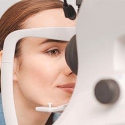
Ophthamological examinations
If you notice changes in central vision , you should have your sight and eyes examined by an ophthalmologist in a clinic.
The ophthalmologist will perform a series of tests to diagnose if you suffer from macular degeneration. Complex eye examinations with the use of specialist equipment is needed.
The examinations may include :
- Vision acuity examination : The test evaluates if the patient can see letters,digits and other signs at different distances, which is very important for reliable monitoring the course of disease and specifying degree of vision damage as well as for legal and insurance purposes..
- Amsler test. Patient is asked to cover one eye, and with the other eye at a net consisting of black straight, vertical and horizontal lines with a dot in the centre . If any of the lines in the net wavy, interrupted or distorted in any way, one may suspect AMD. The test has to be carried out in lenses for hyperopic patients.
Check your sight yourself !
You may perform yourself home the Amsler test, using the presented on this site one. If the patient has a diagnosed AMD, they should perform the test regularly to check if the sight does not worsen.
- Autufluoroscence : Retina pigment epithelium / RPE / is the deepest retina layer. RPE cells are responsible for keeping photoreceptors alive In a dry AMD the RPE layer rarefies causing finally death of the photosensitive cells, which leads to sight worsening. In the advanced stage, the areas of RPE disappearance are huge and called geographical atrophy. Autofluoroscent image is used to describe the size and speed of the area of geographical atrophy increase. It also allows to evaluate druses.
- Fundus examination: Frequently, before this examination, the physician gives the patient drops that are supposed to widen the eye pupils, which allows to examine the bottom of the eye precisely. Next , the ophthalmologist with the use of specialist device watches the “bottom “ of the eye, and looks for different symptoms. of disease. The physician is interested in the presence of druses, retina damages, optic nerve, choroid and blood vessels. For records and better analyses different pictures of fundus are taken both colourful and with special filters, which help in diagnosis.
- Fluorescent angiography : this examination is performed if wet AMD is suspected and to diagnose , among others , blood vessel distortions on fundus. A special pigment is injected into the patients vein, which through blood vessels reaches the retina, detecting vessel damages and their possible locations, or the so- called neovascularisation / CNV /- improper vessels created due to course of wet AMD
- Optical Coherent Eye Tomography / OCT / is a relatively new and totally non-invasive technique used to examine the retina. With the use of OCT, we are able to see the cross-section of retina and its different layers which can be precisely evaluated, for example measure their thickness. It is also possible to see the condition of deeper retina layers by, kind of , looking under its surface and inside. That technique is routinely applied for qualifying to treatment and evaluating retina response to different treatments as well as for precise monitoring progress of disease, which helps to make decisions on starting treatment at the optimal time.
- Tonometry – examination is aimed at measuring pressure inside the eye, excluding all other diseases except the AMD ones. This examination also can also check if applied drops or treatments do not increase pressure, which might be harmful to the eye.
AMD Treatment
Most treatment methods described above concern wet AMD –related to age. It should be stressed that at present there are no specific treatments of dry AMD . However, by applying adequately prepared vitamins and microelements / AREDS/ we can significantly reduce the risk of dry, initial AMD progress into advanced stage or wet form.
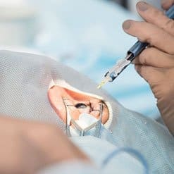
Anti-VEGF drugs
At present, the only registered in Poland and European Union drug in treatment of wet AMD is Lucentis.
Lucentis / international name / ranibizumab
Since loss of sight in wet AMD is caused mainly by the increase of improper blood vessels which finally damage the retina , a drug aimed at stopping the progress of disease. Lucentis is a part of antibody, which binds with the increase factor of vessel endothelium /VEGF / and inhibits its activity. It is believed that VEGF plays a key role in building new blood vessels. Lucentis is injected into vitreous body / vitrous body is a transparent jelly body fulfilling the eye. VEGF is produced continually, therefore injecting of Lucentis should be repeated.
Decision of Lucentis administration is taken by the ophthalmologist, retina specialist , who on the basis of performed diagnostic examinations, determines the frequency and schedule of repeated injections. Most frequently, the patient needs 3 injections as the beginning of therapy every month, and then the decision on next injections is taken depending on progress of disease and how effective the drug is. The medical treatment is not very invasive, however , it requires special preparation before it and monitoring after. The injection into vitreous body should be performed in operating room, which is sterile and aseptic to minimize possible infections or complications.
FACTS:
Lucentis stops or slows down AMD progress, which may lead to blindness. In order to obtain possibly best positive treatment effects, the treatment should be commenced early enough and be repeated as needed.