Lack of eyeball or microphthalmia and prosthetics of eyeball
Table of Content
Inborn anophthalmia – occurs at 1 to 10 cases in 100000 births, which leads to facial bone deformation and serious cosmetic defect. Since it happens to children , it must affect the psyche of little patients, causing different complexes and inhibitions.
The reasons of anophtalmia may be different :
- Primary lack of eyeball or eyeballs takes place when there is no creation of eyeball nucleus
- Secondary – when the damage concerns existing germs
- Successive – when atrophy concerns already created eyeball nucleus.
Hydroxifile expanders
Inborn anophtalmia seriously disturbs facial symmetry. If it is not treated medically, there is no chance for proper prosthetics of orbit and correct setting of eyeball in the future. Technique of hydroxiphile expanders is the latest procedure which allows to reconstruct the orbit and waves in a growing child.
There are many reasons indicating the necessity of orbit prosthetics, which may be divided into primary –resulting from the necessity of enucleating in the course of different eye diseases or inborn lack of eyeball, or secondary- performed even several years before removing the eyeball.
Treatment of anopthalmia
Proper procedure in inborn anopthalmia is , first of all, to assure adequate filling of orbit space. The implant gives the possibility of creating so-called orbit pressure necessary to stimulate development of facial bones, and at the same time, stimulates soft tissues thus assuring the possibility of creating lid crinkles, which are needed for application of thin epiprothesis. Other possible procedures are to make orbitomy or osteotomy. However, such procedures are not performed nowadays due to doubtful cosmetic effects.
Implantation of conformers allows to create tiny crinkles, which needs to be repeated many times.Very good results have been obtained with the implementation of silicon balloons, into which silicon oil was pumped. However, after some time it turned out that the stimulation of orbit was closely connected with the moment of complementing oil, orbit pressure increased in a non-linear way and the procedure had to be repeated frequently.
Lately, more and more popular has become the method of self-expanding hydrophilic implants, which are applied dry into crinkle and by absorbing water, increases gradually, causing soft tissue growth. By producing adequate pressure, development of orbit bone structures is stimulated. There are two basic type: Socket- ensures crinkle development and Orbit- stimulates orbit development. Orbit reconstruction in children ends up with secondary prosthetics and setting a bio-ceramic or hydroxyapatite filling with straight muscle undergrowth in order to obtain a mobile implant, which means a mobile eye prosthesis.
Primary orbit prosthesis
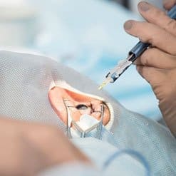
Removal of eyeball is the most drastic surgery in ophthalmology
Unfortunately, diseases like cancer as well as vast injuries, endophtalmity usually leave no choice. In such situations, the metod to be used is removal of eyeball, connected with primary prosthetics or eviscerization together with bio-ceramic implantation filling. It is essential and of ten underestimated in our country to present to the patient possible therapies, different procedures in order to help the patient take the right decision of surgery and post-surgery recommendations. Patients who face eye removal should be supported by psychologists and or psychiatrists. However, the duty of presenting to the patient all the pros and cons oof prosthetics and surgeries.
Indications to enucleation and evisceration of eyeball:
- Non- operative cancer
- Sightless, painful eyeball in glaucoma, which cannot be operated or treated with drugs, including endo-photocoagulation of ciliary body
- Cosmetic / no felling of light /
- Aplasia and hipoplasia
- Prevention of sympathetic inflammation
- Inflammation of eyeball interior In eses with no light feeling, which cannot be treated in standard manner.
- In disturbances of coagulation it is better to perform simultaneous coagulation of retinal artery by laser or diathermy.
Contraindications to evisceration comprise : some cancer diseases, hipoplasia, and aplasia of eyeball in the childhood.
Lack of eyeball leads to partial orbital tissue atrophy, which reaches at least 7%. That is why removal of eyeball or evisceration should be combined with primary orbital prosthesis.
The oldest descriptions of surgery techniques date back to 1885. It is impossible to present all of them, however, it is worth mentioning a very general division of implants:
- Bio-integrating implants, which in time get integrated with patient`s tissue, for example bioceramic, hydroxyapatite, or transplants or dermal fat grafts
- Implants that do nor get biointegrated like silicon, Hema
Each surgery should be performed individually, selecting the best method of procedure.after enucluation and getting control over bleeding, simple muscles are to be spoffed to the implant, and if it is possible in areas of physiological attachments. One of the good methods is to stratify the retina in areas simple muscle attachments and to spoof the whole muscle block to implant. Such procedure allows to avoid many troubles with moving implant and diminish risk of expulsion, obtaining maximal implant mobility. Biointegrating implants are vascularised and filled with connective tissue in about 6 to 9 months.
After such a period, additional titanium hitches may be screwed in to fasten epiprosthesis. Implants that do not get integrated have to be covered with autologous sclera / taken from the patient / broad fascia or artificial material like sheath with Goretex. It is of primary importance that the sheath of eyeball and conjunctiva are closed properly.
Secondary prosthetics
Secondary prosthetics of orbit are surgeries aimed replacing the existing implant for another or implanting after previously performed enucleation. Due to elapsed time since primary surgery, a partial loss of orbital tissues, fibrosis, and difficulties with location identification may be expected. Such surgeries are not easy to perform but possible. After performing primary or secondary orbital prosthesis it happens that additional sub-bone implants are necessary to be placed, which allow proper positioning of epi-prosthesis. Each surgery is completed with fixing a conformer that models the crinkles.
In eyes where crinkle depths got smaller, it is necessary to perform plastic surgery. Sometimes, it is necessary carry out mucous membrane transplantation from oral cavity. Due to limited chances of maintaining hygiene, skin drafts should not be performed. A very promising but expensive technique is to implant hydrophilic Socket expanders , which allow to shape crinkles properly , avoiding difficult transplantation of mucous membrane.
Post eyeball expanders, called PIN expanders are also applied in cases of serious loss of orbital tissue, which need to be supplemented. PIT expanders allow for proper placement of implants in orbit, and it diminishes clearly effect of upper eyelid collapsing and epi-prosthesis.
Complications , which may occur in above mentioned surgeries include, first of all, exposure, migration and expulsion of the implant, very rarely infection or difficult to control hemorrhage.
Prosthesis is completed when proper epi-prosthesis is made and possibly, surgical placement of eyelids, mainly when there is a big asymmetry caused by involution.of orbit tissue or injuries.