Streamlining of lacrimal ducts and nasal-lacrimal ducts
Table of Content
Lacrimal ducts begin with lacrimal points located on lacrimal papilla. Lacrimal points lead to upper and lower lacrimal duct, which in their final run, enter the lacrimal sac either individually or through common duct. Next, nasal-lacrimal duct connects lacrimal sac with nasal cavity, more precisely beneath lower nasal concha.
Structures that may easily get clogged
In children due to :
- Congenital obstruction of nasal ducts / most frequently /
- Clogging of lacrimal ducts caused by different injuries.
- Nasal sept
- Chemical or thermal burns
- History of inflammatory, viral or fungal diseases
- Cancer diseases
In newborns tears begin to appear in a few days up to a few weeks after birth. Symptoms of lacrimal duct clogging appear relatively later.
In adults due to :
- Clogging of lacrimal ducts in adults a result of different injuries
- Nasal septum
- Chemical and thermal burns
- Chronic sinusitis
- History of inflammatory, viral and fungal diseases
- Cancer diseases
- Auto- immunological diseases like sarkoidosis and Wegener granuloma
- Tuberculosis
- Polyps
- Mucocellae.
Symptoms of duct occlusion:
- Tearing / epiphora /
- Pain
- Reddening
- Purulent mucus in conjunctival sac / , often brownish, yellowish and whitish
- Eye area irritation
- Swelling around lacrimal ducts
- Infections are accompanied with fever
The first symptom is usually tearing.
Symptoms may intensify in the course of upper airway inflammation./ cold, sinusitis /.Besides , when one is exposed to wind, cold, rain or sunshine the symptoms become noticeable or intensify.
What increases clogging of lacrimal ducts?
- Premature birth
- Structural and functional problems of lacrimal ducts caused by infections
- Cases of lacrimal ducts obstruction in family history
Treatment:
- Surgery – unique method of laser micro endoscopy
- Anesthesia – in adults –local; in children – general
- Duration – about 20 minuyes
- Dismissal – one to two hours after surgery
Our clinic has been performing surgery of lacrimal ducts since 1992 and so far , about a few thousand of them have been carried out, using different methods.
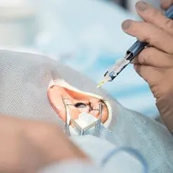
Method of laser micro-endoscopy – consists In inserting a micro-endoscope of 0.4 mm. diameter with a laser optical fiber , which emits light of 980 nm. length into lacrimal sac. The ophthalmological surgeon , during the whole surgery , on the whole length of ducts, is able to evaluate the anatomical condition. When some pathology is diagnosed, the laser turns on and lacrimal ducts are restored. At present ,it is the only surgical method, which allows to perform full reconstruction of lacrimal ducts at any level of obstruction, under control of endoscope .In case of extensive damages, it is possible to perform a typical , classical fistula of lacrimal sac into nasal cavity. The surgery is completed when silicon intubation pipes are inserted, and next removed after 6 to 9 months.

Endoscope used in laser micro-endoscopy
In children –mainly performed is reconstruction of lacrimal ducts / recanalization /w
In adults –fistula between lacrimal sac and nasal cavity /dacryorhinostomia/
In rare cases of total agenesis / inborn lack of lacrimal ducts or complete destruction due to chemical, thermal or mechanical injuries, it is possible to perform an artificial
connection of conjunctive sac to nasal cavity coniuctivodacryocystorhinostomia = CDCR) with implantation of so-called John`s pipe. This surgery is also performed by laser micro-endoscope method, due to which no scars are left on face.
Reconstruction of lacrimal ducts
It is possible to perform by laser micro-endoscopic method and also by retrograde intubation / inserting modified Whorst tube through the healthy canal, backward to damaged canal and removal of adhesions and scars with laser or classical surgery. When both canals are damaged, it is possible to reconstruct them by our own method from the side of lacrimal sac or common canal.
Treatment of fungal inflammations and Actinomycetes.
Treatment consists in surgical removal of fungal mass or Actinomycetes, deposits and next pharmacological treatment .Most frequently, it is performed through natural canal holes , not breaking continuity of canal walls and leaving no scars both on skin and canal wall.. In case of huge deposits , it is sometimes necessary to perform canaliculotomia / cutting canal wall and extracting deposits.
Treatment of children.
In our clinic, we recommend that children should undergo probing before spreaders reach area of lacrimal sac / up to 3-6 months of age /. In case of already existing spreaders and recesses of lachrymal sacs and dacryomycopyocels / muco purulent cysts / , we suggest carrying out recanalization of lacrimal ducts even in children under one year of age.
Post – surgery recommendations:
- Regaining normal lifestyle – usually 12 hours after surgery
- Check up –after one month post surgery
Complications (very rare)
- Lid swelling
- Subcutaneous hematoma
- Slight bleeding from nose
- Damages of canals caused by not abiding recommendations
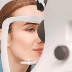
Both eyes are examined
Consultation with Mr. Thorsten Meyer
Both eyes are examined
Both eyes are examined
Both eyes are examined
Both eyes are examined
Both eyes are examined
Both eyes are examined
Both eyes are examined
Both eyes are examined
Both eyes are examined
Both eyes are examined
Both eyes are examined
Both eyes are examined
Both eyes are examined
Both eyes are examined
With general anesthesia
With general anesthesia