Procedure of removing tear duct and nasal lacrimal obstructions
Table of Content
Tear ducts begin with lacrimals points lying on the lacrimal papillae. Lacrimal points lead to upper and lower tubular tear which in its final run flows into the lacrimal sac, separately or through a common duct. Then the nasal-lacrimal duct connects the lacrimal sac with the nasal cavity, closely below the lower nasal concha.
Structures that can be easily clogged
Structures that may easily get clogged:
In children as a result of:
- Congenial obstruction of the nasal passages (most frequently)
- Clogging of the lacrimal ducts due to various injuries
- Deviation of the nasal septum
- Chemical or thermal burns
- Chronic sinusitis
- Viral and fungal inflammations
- Cancer
In neonates, tears start to be produced within a few days after birth, up to several weeks thereafter. Likewise , the symptoms of the lacrimal ducts obstruction appear later.
In adults as a result of :
- Mostly, due to different types of injuries
- Deviations of the nasal septum
- Chemical and thermal burns
- Chronic sinusitis
- Viral and fungal inflammations
- Cancer
- Auto-immuno diseases such as sarcoidosis, Wegner’s granulomatosis
- Tuberculosis
- Polyps
- Mucocellae.
Symptoms of lacrimal duct obstruction:
- Tearing (epiphora).
- Pain
- Redness
- Muco-purulent secretions in the eye (conjunctival sac), often – yellow-white
- Irritation of the eye area
- Swelling around tear ducts
- Infections are often accompanied by fever
The first symptom is mostly lacrimation.
Symptoms may intensify in the course of inflammation of the upper respiratory tracts (colds, sinusitis). In addition, during exposure to wind, cold and sunlight, symptoms may become noticeable or may intensify.
What increases the risk of blocking the tear ducts?
- Premature birth
- Structural and functional problems with lacrimal ducts caused by infections
- Family history of lacrimal duct obstructions
Treatment:
- Surgical – a unique method of laser micro endoscopy
- Anesthesia: in adults- under local anesthesia; in children- in general anasthesia
- It takes about 20 min.
- Discharge from hospital –about 1 to 2 hours after surgery
Lacrimal duct surgery has been performed by our team since 1992. Up till now, we have conducted several thousand treatments of lacrimal duct patency restoration by all methods.
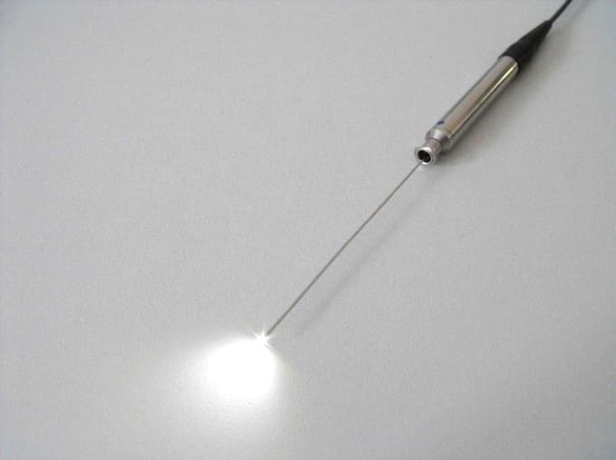
Micro-endoscopic laser method consists in entering a micro-endoscope of 0.4 mm in diameter with fiber optic laser emitting light of 980 nm long into the lacrimal duct .Ophthalmologist surgeon throughout the whole surgery on the entire length of lacrimal duct is able to assess the anatomical status. Upon the detection of the pathology, the laser is activated and lacrimal ducts are reconstructed .At present, this is the only surgical method ,which allows to make a complete reconstruction of the lacrimal duct obstruction at any level, under the control of the endoscope. In the event of extensive damage, the surgeon can make a classical fistula of lacrimal sac into the nasal cavity. The treatment ends up with entering silicone intubation tubes, which are removed after 6-9 months.

One of the endoscopes used in laser micro-endoscopy surgery
In children, it is mainly performed in reconstruction of lacrimal ducts (recanalization).
In adults – in lacrimal sac fistula of the nasal cavity (dacryorhinostomia).
In rare cases of total agenesis (congenital absence) of tear ducts or total destruction as a result of chemical, thermal or mechanical injuries it is possible to carry out artificial connection of conjunctival sac to the nasal cavity (coniuctivodacryocystorhinostomy = CDCR) with implantation of so-called Johns tube. This procedure is performed also by micro-endoscopic laser. and there are no scars left on the face.
Reconstruction of the lacrimal ducts.
It is possible to carry out the reconstruction by a micro-endoscopic-laser, as well as by retrograde intubation (placing a modified Whosrt probe through healthy duct backward to the duct that is damaged, removing the adhesions or scars by laser or classically-surgically.
In case of both ducts damaged , they can be reconstructed by our own method, from the side of lacrimal sac or common duct)
Treatment of fungal dermatitis, and Actinomycetes.
Treatment of fungal inflammations and Actinomycetes inflammation consists in surgical removal of the fungus mass or Actinomycetes deposits , and subsequent pharmacological treatment. Treatment is most often conducted through natural duct openings without affecting the continuity of tubular wall and leaving no scars either on skin or on the wall of the duct . when there are huge deposits it is sometimes necessary to perform canaliculotomy (duct wall incision and extraction of deposits).
Treatment of childrenAt our center, we recommend lacrimal duct probing in children before ectasis reach lacrimal sac (up to 3-6 months of age). In the case of existing ectasis and lacrimal sac recesses and the innate dacryomycopyocele (muco-purulent cyst), we offer recanalization of the lacrimal ducts in children, even below 1 year of age.
Recommandations after surgery:
- Return to normal life takes place usually up to 12 hours after surgery
- Check up is in one month after surgery
Complications (very rare):
- Swelling of eyelids
- Subcutaneous hematoma
- Slight sub-bleeding from nose
- Damage of ducts caused by non-compliance with recommendation
Occluded tubules in children
Quite a common problem in children is closed nasolacrimal duct, usually causing tearing and eye suppuration . In very young children, it is recommended to massage the area. If there is no spontaneous patency or the child’s condition suggests such a need, probing of the lacrimal duct should be carried out (this treatment in our center is performed under local anesthesia). If it does not give a positive effect, micro –endoscopy of lacrimal ducts is to be considered, and possibly simultaneous laser treatment.
Post-traumatic occlusion of tubules
A common result of road accidents is damage of lacrimal ducts . This problem can be solved only by reconstruction of ducts and, in extreme cases, by CDCR surgery (at our center also performed by using laser techniques that do not leave scars), involving the implantation of a silicone tube that permanently leads out tears.
Excessive tearing
Excessive tearing may also have another origin, besides obstruction like: excessive laxity of the eyelids, incorrect lacrimal points` setting, abnormal shape and size of the lacrimal duct warts, lacrimal pump dysfunction, vision defects, allergies, etc.
To improve the running of tears, plastic surgeries on the eyelids are necessary, which are also performed at our center.
Persistent purulence
It can also be caused by obstructive lacrimal ducts . It is not possible to achieve any improvement and permanent cure if the lacrimal ducts do not get patent Frequent administration of antibiotics can lead to conjunctival sac and lacrimal duct fungal inflammation
Such a condition requires prompt surgical intervention as well as general and local treatment.
Both eyes are examined
Consultation with Mr. Thorsten Meyer
Covers whole treatment period (6–9 months)
With general anesthesia
With general anesthesia


